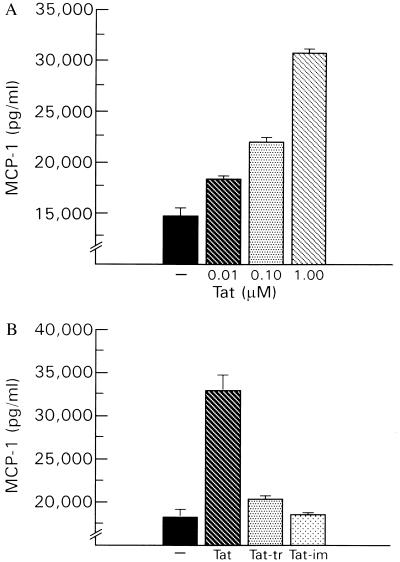
Figure 2.
MCP-1 ELISA analysis of supernatants from variously treated human astrocytes. (A) Astrocytes were grown to near confluency in 35-cm plates. Each well contained 106 cells in 1 ml of medium. The medium was then changed and astrocytes were stimulated with exogenous Tat in doses ranging from 0.01 to 1.0 μM. Twenty hours later, samples were taken for analysis by immunoassay (R&D Systems). As compared with untreated astrocytes (−) that, when grown in tissue culture, express MCP-1 in the absence of stimulation, Tat-stimulated astrocytes showed a significant increase in MCP-1 release. Data are shown as mean + SE for three replicates. (B) Similar experiment except that astrocytes were stimulated with 100 nM of Tat or with an equivalent amount of Tat that had first been either digested with trypsin (Tat-tr) or immunoabsorbed (Tat-im).

