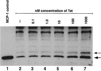
Figure 3.
Western blot analysis of MCP-1 in astrocyte supernatants. In lanes 2–7, 50 μg of protein from variously treated astrocyte supernatants were run on a 15% Tris⋅glycine denaturing gel. Three nanograms of non-glycosylated recombinant MCP-1 (R&D Systems) was run in lane 1 as a control. After protein transfer to nitrocellulose, the blot was probed with a polyclonal antibody that recognizes human MCP-1 (R&D Systems). After washing, an appropriate secondary antibody was applied [horseradish peroxidase conjugated anti-goat (Santa Cruz Biotechnology)] and electrochemiluminescence (Amersham) was used to visualize the bands. The two bands, which are specifically increased in association with Tat, are indicated by arrows. The lower arrow represents a band that runs with an apparent molecular mass of 9 kDa, whereas the upper band, of slightly higher molecular mass, is likely to represent MCP-1 that has been altered by the addition of O-linked carbohydrates. Both forms of MCP-1 are active in vitro (15).

