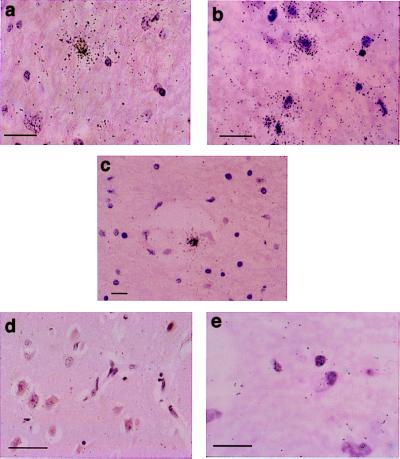
Figure 5.
Detection of MCP-1 RNA in brain tissue by in situ hybridization. Tissue sections were hybridized with an MCP-1 antisense probe. (a and b) Cells within the cerebral white matter of a HIVD patient show a strong signal for the presence of MCP-1 RNA. (c) Signal-positive cells are also seen in perivascular regions. A representative area from a normal patient shows absence of signal (d), as does a sample from an HIVD patient that was hybridized with an MCP-1 sense probe (e). (Scale bars represent 25 μm.)

