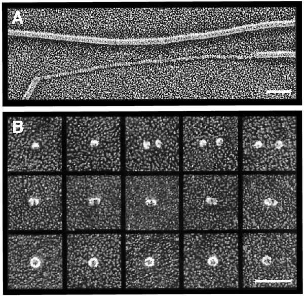
Figure 3.

Deep-etch electron microscopy. (A) Platinum replica of purified P pili incubated in 50% glycerol showing unraveling of one of the pilus rods. (Bar = 50 nm.) (B) Montage of platinum replicas of purified PapC showing putative PapC monomers (top row), dimers (middle row), and ring-shaped complexes (bottom row). The ring-shaped complexes contain central pores 2–3 nm in diameter. (Bar = 50 nm.)
