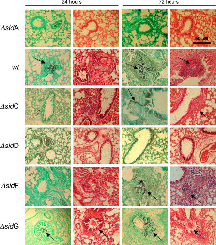
Figure 9. Histopathological Analysis of Aspergillus-Infected Murine Lung Sections.
Comparative histopathology of neutropenic murine lung sections following infection with A. fumigatus wt or A. fumigatus ΔsidA, ΔsidC, ΔsidD, ΔsidF, and ΔsidG mutants. Sections were sampled at 24 and 72 h post-infection, fixed in 4% v/v formaldehyde, and stained using Grocotts Methanamine Silver (GMS), or hematoxylin and eosin (HE). Infectious foci containing fungal hyphae and inflammatory lesions are indicated by arrows over GMS and HE sections, respectively.

