
Fig. 1.
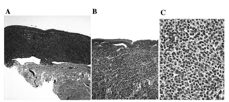
Photomicrographs showing well-demarcated MALT lymphoma infiltration in the substantia propria beneath or invading the conjunctival epithelium in Case 1 (A) and Case 3 (B); higher magnification showing cytologic features of MALT lymphoma (C) (hematoxylin & eosin, original magnification: A, × 50; B, × 100; C, × 400)