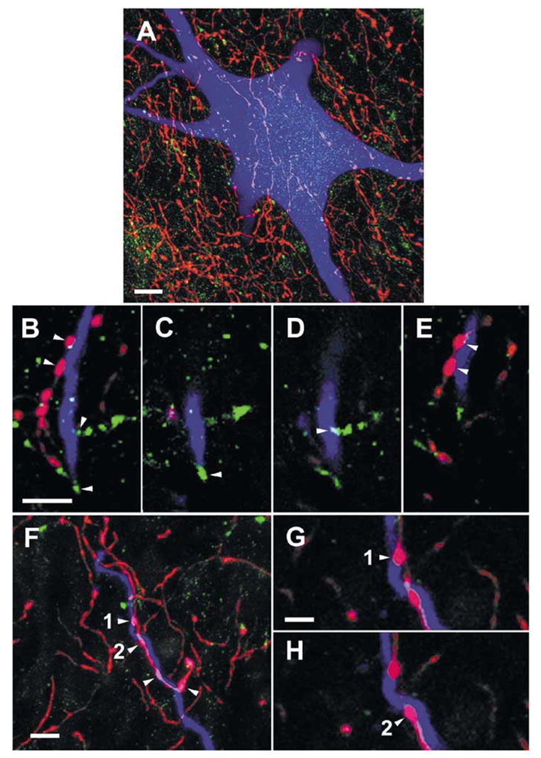
Fig. 7.

Examples of contacts between monoaminergic fibres and commissural interneurons. (A) A projected image compiled from 85 × 0.5-μm optical sections through the soma of a cell (with RF input; cell 6 in Table 1, records in Fig. 6G-I), showing the abundance of 5-HT-immunoreactive axons bearing varicosities (red) in the vicinity of the cell soma and the relative infrequency of d.b.h.-immunoreactive axons (green) in the same area. (B) A projected image (compiled from 26 × 0.5-μm optical sections) of a dendrite from the cell shown in A. Boutons immunoreactive for 5-HT (shown in red) and d.b.h. (green) can be seen close to the dendrite. (C–E). Single optical sections from the series shown in B illustrate contacts made by the boutons labelled by arrowheads in B onto the dendrite. (F) A further projected image (compiled from 16 × 0.5-μm optical sections) showing a 5-HT-containing axon forming four boutons along a labelled dendrite (originating from a cell with input from both RF and group II afferents; cell 4 in Table 1). (G) and (H) show the terminals labelled 1 and 2 in (F) at higher magnification. Scale bars, 10 μm (A), 5 μm (B–F), 2.5 μm (G–H).
