Abstract
Serial image guided 31P magnetic resonance spectroscopy (MRS) studies were performed in eight patients with non-Hodgkin's lymphoma to determine the changes in phosphorus metabolites that occur in vivo in response to chemotherapy. Pre-treatment spectral characteristics were different in high and low grade lymphoma. A larger inorganic phosphate (Pi) peak was seen in high grade NHL relative to phosphomonoesters (PME) or beta adenosine triphosphate (beta ATP), producing significant differences in the PME/Pi and Pi/beta ATP metabolite ratios, and probably reflecting a larger hypoxic cell fraction within the high grade lymphomas. Consistent metabolite changes were seen with treatment, and before reductions in tumour bulk had occurred. Alterations in tumour energetics with changes in Pi and beta ATP, and increases in phospholipid turnover reflected as an increase in the phosphodiester (PDE) resonance were detected. Changes were seen between days 10 and 27 in low grade lymphoma treated with oral alkylating therapy and between days 1 and 5 in lymphoma treated with intensive combination chemotherapy. Increases in the PDE/beta ATP metabolite ratio may be an early indicator of response to chemotherapy in human tumours. These studies illustrate the feasibility and clinical potential of image guided 31P MRS as a means of assessing response to therapy.
Full text
PDF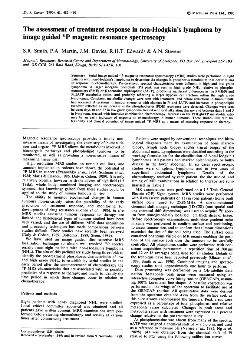
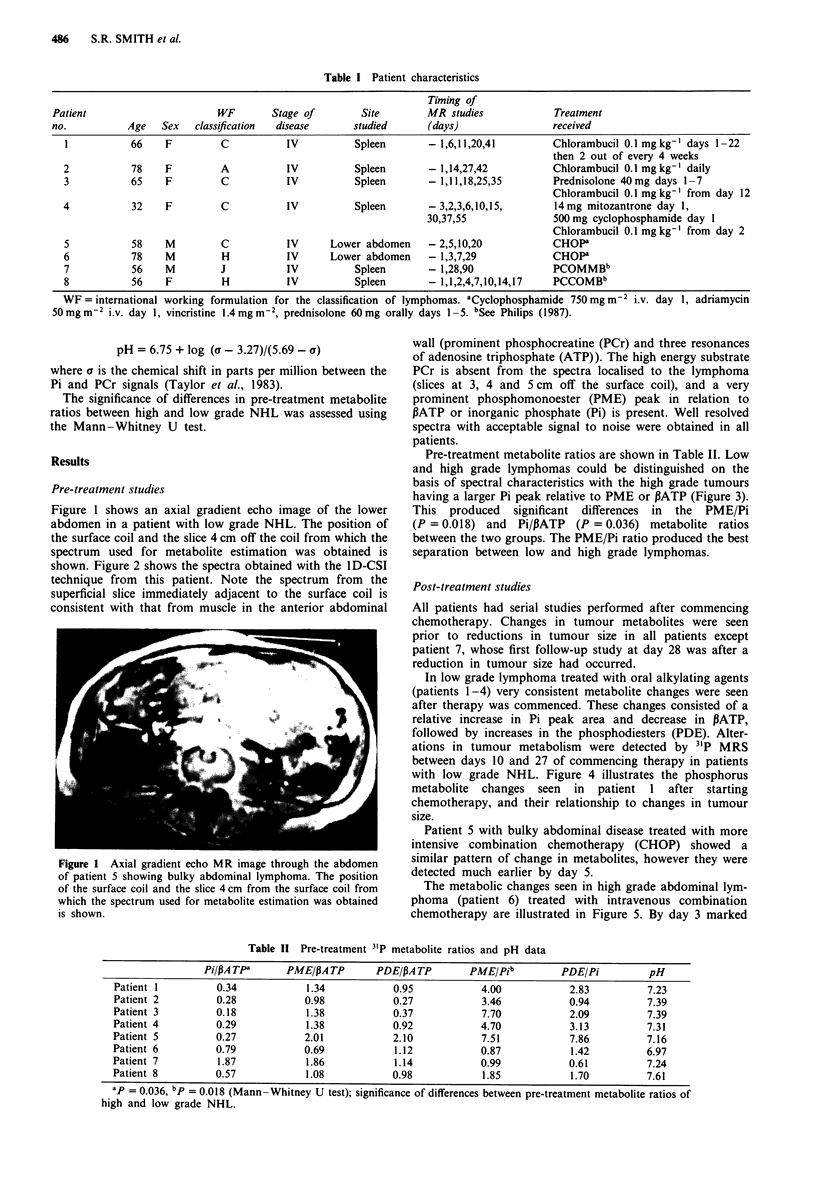
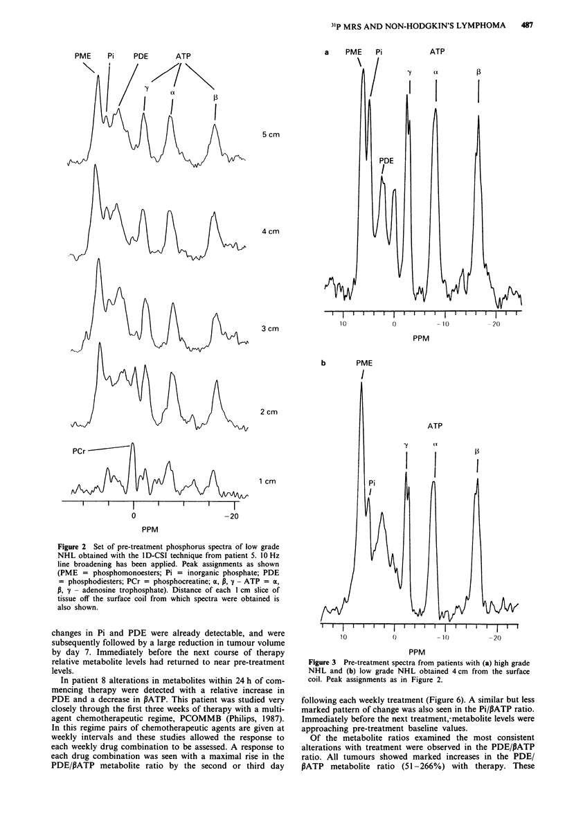
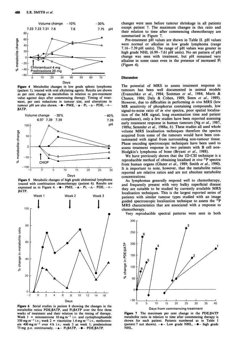
Images in this article
Selected References
These references are in PubMed. This may not be the complete list of references from this article.
- Bottomley P. A. Human in vivo NMR spectroscopy in diagnostic medicine: clinical tool or research probe? Radiology. 1989 Jan;170(1 Pt 1):1–15. doi: 10.1148/radiology.170.1.2642336. [DOI] [PubMed] [Google Scholar]
- Bryant D. J., Bydder G. M., Case H. A., Collins A. G., Cox I. J., Makepeace A., Pennock J. M. Use of phosphorus-31 MR spectroscopy to monitor response to chemotherapy in non-Hodgkin lymphoma. J Comput Assist Tomogr. 1988 Sep-Oct;12(5):770–774. doi: 10.1097/00004728-198809010-00010. [DOI] [PubMed] [Google Scholar]
- Cohen J. S., Lyon R. C., Chen C., Faustino P. J., Batist G., Shoemaker M., Rubalcaba E., Cowan K. H. Differences in phosphate metabolite levels in drug-sensitive and -resistant human breast cancer cell lines determined by 31P magnetic resonance spectroscopy. Cancer Res. 1986 Aug;46(8):4087–4090. [PubMed] [Google Scholar]
- Daly P. F., Cohen J. S. Magnetic resonance spectroscopy of tumors and potential in vivo clinical applications: a review. Cancer Res. 1989 Feb 15;49(4):770–779. [PubMed] [Google Scholar]
- Daly P. F., Lyon R. C., Faustino P. J., Cohen J. S. Phospholipid metabolism in cancer cells monitored by 31P NMR spectroscopy. J Biol Chem. 1987 Nov 5;262(31):14875–14878. [PubMed] [Google Scholar]
- Evanochko W. T., Ng T. C., Glickson J. D. Application of in vivo NMR spectroscopy to cancer. Magn Reson Med. 1984 Dec;1(4):508–534. doi: 10.1002/mrm.1910010410. [DOI] [PubMed] [Google Scholar]
- Glaholm J., Leach M. O., Collins D. J., Mansi J., Sharp J. C., Madden A., Smith I. E., McCready V. R. In-vivo 31P magnetic resonance spectroscopy for monitoring treatment response in breast cancer. Lancet. 1989 Jun 10;1(8650):1326–1327. doi: 10.1016/s0140-6736(89)92717-7. [DOI] [PubMed] [Google Scholar]
- Glazer G. M., Smith S. R., Chenevert T. L., Martin P. A., Stevens A. N., Edwards R. H. Image localized 31P magnetic resonance spectroscopy of the human liver. NMR Biomed. 1989 Apr;1(4):184–189. doi: 10.1002/nbm.1940010406. [DOI] [PubMed] [Google Scholar]
- Griffiths J. R., Cady E., Edwards R. H., McCready V. R., Wilkie D. R., Wiltshaw E. 31P-NMR studies of a human tumour in situ. Lancet. 1983 Jun 25;1(8339):1435–1436. doi: 10.1016/s0140-6736(83)92375-9. [DOI] [PubMed] [Google Scholar]
- Hesketh T. R., Moore J. P., Morris J. D., Taylor M. V., Rogers J., Smith G. A., Metcalfe J. C. A common sequence of calcium and pH signals in the mitogenic stimulation of eukaryotic cells. Nature. 1985 Feb 7;313(6002):481–484. doi: 10.1038/313481a0. [DOI] [PubMed] [Google Scholar]
- Naruse S., Hirakawa K., Horikawa Y., Tanaka C., Higuchi T., Ueda S., Nishikawa H., Watari H. Measurements of in vivo 31P nuclear magnetic resonance spectra in neuroectodermal tumors for the evaluation of the effects of chemotherapy. Cancer Res. 1985 Jun;45(6):2429–2433. [PubMed] [Google Scholar]
- Ng T. C., Grundfest S., Vijayakumar S., Baldwin N. J., Majors A. W., Karalis I., Meaney T. F., Shin K. H., Thomas F. J., Tubbs R. Therapeutic response of breast carcinoma monitored by 31P MRS in situ. Magn Reson Med. 1989 Apr;10(1):125–134. doi: 10.1002/mrm.1910100112. [DOI] [PubMed] [Google Scholar]
- Ng T. C., Majors A. W., Vijayakumar S., Baldwin N. J., Thomas F. J., Koumoundouros I., Taylor M. E., Grundfest S. F., Meaney T. F., Tubbs R. R. Human neoplasm pH and response to radiation therapy: P-31 MR spectroscopy studies in situ. Radiology. 1989 Mar;170(3 Pt 1):875–878. doi: 10.1148/radiology.170.3.2916046. [DOI] [PubMed] [Google Scholar]
- Ng T. C., Vijayakumar S., Majors A. W., Thomas F. J., Meaney T. F., Baldwin N. J. Response of a non-Hodgkin lymphoma to 60Co therapy monitored by 31P MRS in situ. Int J Radiat Oncol Biol Phys. 1987 Oct;13(10):1545–1551. doi: 10.1016/0360-3016(87)90323-3. [DOI] [PubMed] [Google Scholar]
- Oberhaensli R. D., Hilton-Jones D., Bore P. J., Hands L. J., Rampling R. P., Radda G. K. Biochemical investigation of human tumours in vivo with phosphorus-31 magnetic resonance spectroscopy. Lancet. 1986 Jul 5;2(8497):8–11. doi: 10.1016/s0140-6736(86)92558-4. [DOI] [PubMed] [Google Scholar]
- Schuldiner S., Rozengurt E. Na+/H+ antiport in Swiss 3T3 cells: mitogenic stimulation leads to cytoplasmic alkalinization. Proc Natl Acad Sci U S A. 1982 Dec;79(24):7778–7782. doi: 10.1073/pnas.79.24.7778. [DOI] [PMC free article] [PubMed] [Google Scholar]
- Semmler W., Gademann G., Bachert-Baumann P., Zabel H. J., Lorenz W. J., van Kaick G. Monitoring human tumor response to therapy by means of P-31 MR spectroscopy. Radiology. 1988 Feb;166(2):533–539. doi: 10.1148/radiology.166.2.3336731. [DOI] [PubMed] [Google Scholar]
- Semmler W., Gademann G., Schlag P., Bachert-Baumann P., Zabel H. J., Lorenz W. J., von Kaick G. Impact of hyperthermic regional perfusion therapy on cell metabolism of malignant melanoma monitored by 31P MR spectroscopy. Magn Reson Imaging. 1988 May-Jun;6(3):335–340. doi: 10.1016/0730-725x(88)90410-9. [DOI] [PubMed] [Google Scholar]
- Sostman H. D., Armitage I. M., Fischer J. J. NMR in cancer. I. High resolution spectroscopy of tumors. Magn Reson Imaging. 1984;2(4):265–278. doi: 10.1016/0730-725x(84)90192-9. [DOI] [PubMed] [Google Scholar]
- Steen R. G. Response of solid tumors to chemotherapy monitored by in vivo 31P nuclear magnetic resonance spectroscopy: a review. Cancer Res. 1989 Aug 1;49(15):4075–4085. [PubMed] [Google Scholar]
- Taylor D. J., Bore P. J., Styles P., Gadian D. G., Radda G. K. Bioenergetics of intact human muscle. A 31P nuclear magnetic resonance study. Mol Biol Med. 1983 Jul;1(1):77–94. [PubMed] [Google Scholar]
- Wehrle J. P., Glickson J. D. 31P NMR spectroscopy of tumors in vivo. Cancer Biochem Biophys. 1986 Jul;8(3):157–166. [PubMed] [Google Scholar]


