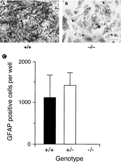
Figure 2.
Failure of LIFR−/− neural cells to express GFAP. Neuroepithelial cells from E12 forebrain were plated in vitro at a density of 2.5 × 10⁴ per 200 mm2 into multiwell plates (Falcon 3047) and cultured for 20 days in the presence of serum. Cultures then were stained by immunoperoxidase for the presence of GFAP. The LIFR+/+ cultures contained large numbers of GFAP-positive cells (A and C) whereas the LIFR−/− cultures (B and C) contained few if any (<2 cells per well in every case). The data shown in C are the mean and SEM obtained from three separate experiments, each with three replicates.

