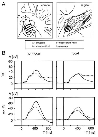
Figure 1.
Area of electrode locations at which maximal AMTL-N400s to words were recorded and grand averages of AMTL-N400s. (A) Schematics of recording sites of AMTL-N400s to words. Hatched area indicates the area in which maximal AMTL-N400 potentials were recorded in all patients. (B) Grand averages of AMTL-N400s in the nonfocal and focal temporal lobe in patients with extrahippocampal lesions and without hippocampal sclerosis (n = 21) as well as in patients with hippocampal sclerosis (n = 29). HS, hippocampal sclerosis. Solid line, first presentations; dashed line, word repetitions.

