Figure 1.
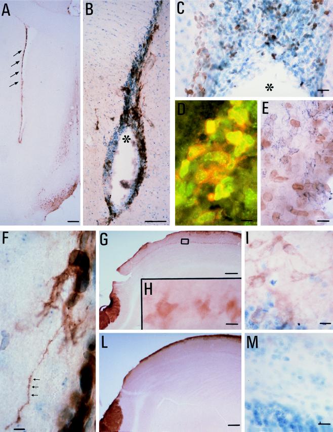
(A and B) p75LNGFR immunoreactivity is localized in the SVZ of the adult rat brain at rostrocaudal level 1.7 mm (refs. 13 and 14, tyramide-intensification procedure). The arrows point to positive staining in the SVZ, and the asterisk indicates the lateral ventricle. In the dorso-lateral corner of the lateral ventricle sampled at this level, numerous cycling, Ki67-positive cells (C, avidin-biotin technique; the asterisk indicates the ventricle), partially coexisting with p75LNGFR and neural cell adhesion molecule positivity are observed. (D) Double-labeling immunofluorescence, in which the green fluorescence refers to Ki67-positive nuclei and red fluorescence refers to p75LNGFR. Fluoresceine isothiocyanate- and rhodamine-conjugated Ig were from Dako. (E) Double-labeling avidin-biotin technique. The brown diaminobenzidine product refers to Ki67 positivity, whereas the blue chloronaphthol product refers to neural cell adhesion molecule positivity. Fibers departing from p75LNGF-positive elements are observed in the SVZ of the lateral ventricle during EAE (F). TrkA positivity also was observed in the mitral cells in the olfactory bulb (G: low-power magnification, 2×; H: higher magnification, 40×, referring to the square area indicated in G; I: toluidine, controstained TrkA-positive section). TrkA positivity was not observed in the mitral cells of control rats (L and M). [Bars = 200 μm (A, G, and L), 50 μm (B), 25 μm (C, F, H, I, and M), and 15 μm (D and E).]

