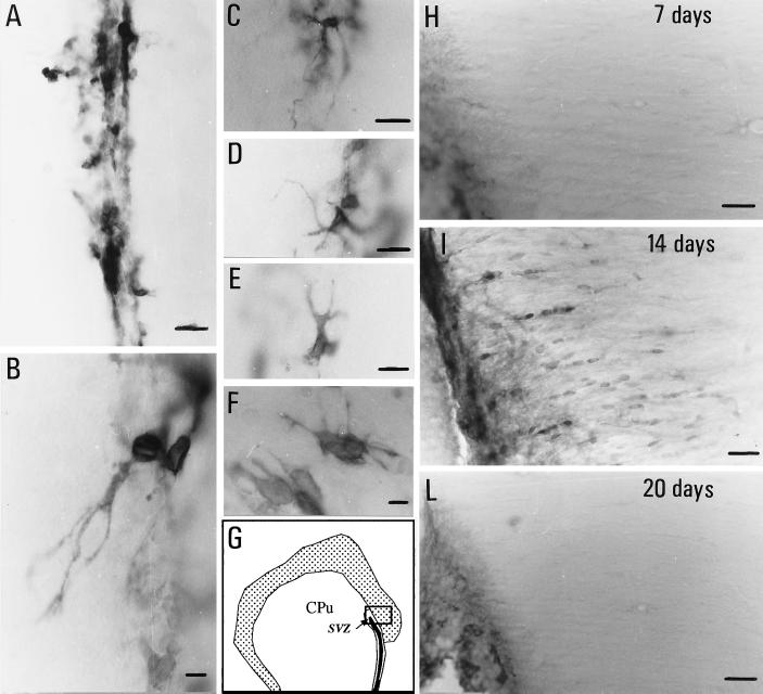
Figure 3.
p75LNGFR immunoreactivity in the SVZ of control (A) and EAE (B–F) rats, 21 days after immunization. Micrographs B–F illustrate the diffuse and branched fiber outgrowth observed during EAE. G shows the area sampled by micrographs H–L. Migrograph I shows p75LNGFR-positive cells in the corpus callosum 14 days after immunization, which were not observed at other time-points during EAE nor in control rats. CPu, caudato-putamen nucleus. (Bars = 25 μm.)

