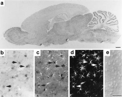
Figure 5.
In situ hybridization histochemistry for NAALADase-like mRNAs and colocalization with GFAP. Expression of NAALADase-like mRNA visualized with cRNA probes appears to be widespread in the brain, with midbrain-brainstem structures generally exhibiting higher hybridization than forebrain areas, except for the olfactory bulb, which shows a relatively high level of expression (a). Note particularly intense labeling of a narrow band near the Purkinje cell layer of the cerebellum. The colocalization of NAALADase-like mRNA to GFAP-positive astroglia in a field of rat hippocampal cells in a coronal brain section (arrows indicate examples of dual-labeled cells in registry. (b) Labeling with a NAALADase cRNA probe shows that small cell bodies in the hippocampus are encircled with dark staining for NAALADase-like message. (d) Immunostaining of this same group of hippocampal cells for the astrocyte-specific marker GFAP is shown, indicated by fluorescent fluorescein isothiocyanate-labeling of fibers within the cell processes. (c) A double-exposure photomicrograph shows that the two signals colocalize to the same cells, with fluorescent GFAP-positive processes extending from dark perinuclear halos of NAALADase labeling. (e) Bright-field photomicrograph of an adjacent section of tissue to which a control sense NAALADase cRNA was applied shows no labeling. [Bars = 50 μm (a) and 5 μm (b–e).]

