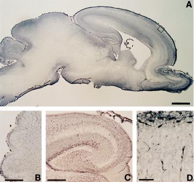
Figure 1.
Reelin (G10)-immunostaining in the neonatal [postnatal day 0 (P0)] rat. Low magnification of a brain sagittal section (A) shows that Reelin is most abundant in the marginal zone of the cerebral cortex, hippocampus, cerebellum, and olfactory bulb. Higher magnification of the cerebellum (B) shows that Reelin is most abundant in the EGL. C shows that the highest Reelin expression is around the hippocampal fissure. D, which is a high magnification of the cortical plate boxed in A, shows that Reelin is highly expressed in large horizontally oriented fusiform neurons, presumably CR cells. [Bars = 1 mm (A), 25 μm (B and C), and 50 μm (D).]

