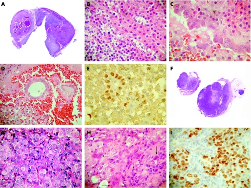
Figure 1 (A) Nodular hyperplasia of the left superior parathyroid gland with areas of haemorrhage and cystic degeneration. (B) Chief cells and oxyphil cells in the hyperplastic left inferior parathyroid gland. (C) Left superior parathyroid gland cyst lined by malignant cells contrasting with bland chief/oxyphil cells in the wall of the cyst. (D) Malignant glands within the left superior parathyroid gland cyst. (E) Malignant glands within left superior parathyroid cyst showing TTF‐1 nuclear staining. (F) Section of lymph node replaced by metastatic carcinoma. (G) Sheet of carcinoma cells within the lymph node with occasional gland‐like spaces. (H) Metastatic carcinoma with osteoclastic giant cells (arrows) within lymph node. (I) Metastatic carcinoma within lymph node showing TTF‐1 nuclear staining.

An official website of the United States government
Here's how you know
Official websites use .gov
A
.gov website belongs to an official
government organization in the United States.
Secure .gov websites use HTTPS
A lock (
) or https:// means you've safely
connected to the .gov website. Share sensitive
information only on official, secure websites.
