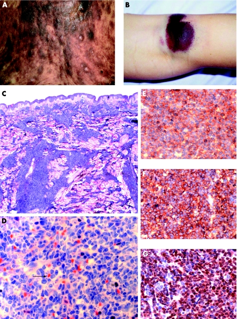
Figure 1 Morphological and immunophenotypic features of CD4+/CD56+ haematodermic neoplasm (group 1). (A) The trunk of a patient with generalised red to violet‐brown plaques and nodules (case 6). (B) Bruise‐like lesion on the right knee (case 3). (C and D) Histological findings of skin biopsy show dense infiltration of medium‐sized blastoid tumour cells without angiocentric growth pattern, and frequent atypical mitoses (solid arrow) and massive erythrocyte extravasation (open arrowhead). (E) Strong membranous staining of CD56. (F) Strong membranous staining of CD123. (G) Strong nuclear staining of TdT.

An official website of the United States government
Here's how you know
Official websites use .gov
A
.gov website belongs to an official
government organization in the United States.
Secure .gov websites use HTTPS
A lock (
) or https:// means you've safely
connected to the .gov website. Share sensitive
information only on official, secure websites.
