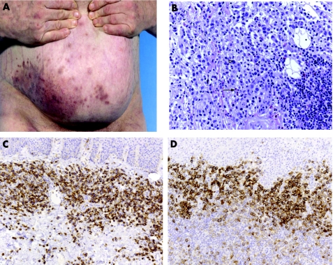
Figure 4 Morphological and immunophenotypic features of lymphomatoid papulosis with co‐expression of CD56. (A) Grouped papules on the right abdomen (case 30). (B) Histological findings are large atypical lymphocytes (open arrowhead) embedded in a mixed inflammatory infiltrate with eosiniophilic granulocytes (closed arrow). (C) Immunohistology revealed expression of CD30. (D) Immunohistology revealed expression of CD56.

An official website of the United States government
Here's how you know
Official websites use .gov
A
.gov website belongs to an official
government organization in the United States.
Secure .gov websites use HTTPS
A lock (
) or https:// means you've safely
connected to the .gov website. Share sensitive
information only on official, secure websites.
