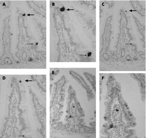
Figure 5 Sequential 5 μm section immunohistochemistry with anti‐human defensin (HD)‐5 antibody (A, B), anti‐lysozyme antibody (C, D) and anti‐intestinal trefoil factor antibody (E, F) seen at low (A, C, E) and high (B, D, F) magnification. An intermediate cell on the villus tip (large arrow) is seen to express HD‐5 and lysozyme, whereas an intermediate cell towards the villus base expresses HD‐5 but not lysozyme (small arrow). The lack of Paneth cell staining using the rabbit polyclonal anti‐intestinal trefoil factor antibody (E, F) in these sequential sections also serves as a control for the HD‐5 and lysozyme polyclonal antisera.

An official website of the United States government
Here's how you know
Official websites use .gov
A
.gov website belongs to an official
government organization in the United States.
Secure .gov websites use HTTPS
A lock (
) or https:// means you've safely
connected to the .gov website. Share sensitive
information only on official, secure websites.
