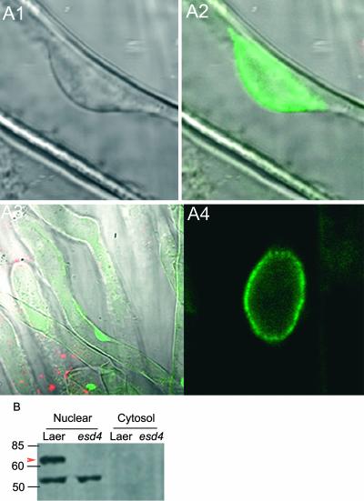
Figure 6.
Subcellular Localization of the ESD4:GFP Fusion Protein.
(A) Confocal microscopy images of the location of ESD4:GFP in esd4-1 mutants carrying the 35S::ESD4:GFP transgene. ESD4:GFP, which complements the esd4-1 mutation, is localized predominantly to the periphery of the nucleus.
(A1) Transmissible light image of the nucleus of a root hair cell.
(A2) The nucleus shown in (A1), with the image collected in the 500- to 530-nm range. The ESD4:GFP signal is located at the periphery of the nucleus.
(A3) The root hair cell shown in (A1) and (A2) at lower magnification, with the image collected in the 500- to 530-nm range. ESD4:GFP is located in the nucleus.
(A4) Nucleus of a root epidermal cell, with the image collected in the 500- to 530-nm range. The ESD4:GFP signal is located at the periphery of the nucleus.
(B) Detection of ESD4 protein in nuclear extracts. Nuclear and cytosolic proteins from 10-day-old seedlings of Ler and esd4-1 were probed with an antibody against ESD4. The arrowhead indicates the candidate ESD4 protein of the expected size (65 kD) that is absent in esd4. The smaller protein is presumed to represent cross-reaction of the antibody.

