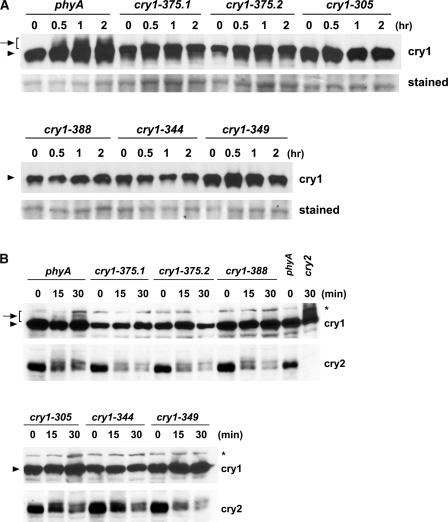
Figure 4.
Immunoblots Showing the Lack of cry1 Phosphorylation and Normal cry2 Phosphorylation in Different cry1 Mutant Alleles.
(A) Samples were prepared from etiolated seedlings exposed to 25 μmol·m−2·s−1 blue light for the time indicated or from samples kept in the dark (0 h), and immunoblots were probed with anti-CRY1 antibodies (cry1). For a loading control, the membrane was stained with Ponceau red, and a portion of the stained blot showing unspecified proteins is included (stained).
(B) Samples were prepared from etiolated seedlings exposed to 7 μmol·m−2·s−1 blue light for the time indicated or from samples kept in the dark (0 min). The immunoblot was probed first with anti-CRY2 antibody (cry2), stripped, and reprobed with anti-CRY1 antibody (cry1).

