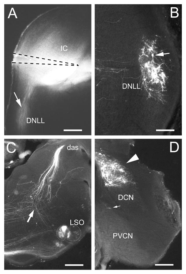
Figure 1.

A. Epifluorescent image of coronal section through midlevel of IC from P7 ferret kit showing position of DiI-pin. Note that the brightest DiI-labeling filled the ventromedial part of the central nucleus of the IC. Retrogradely labeled fibers are visible in the ipsilateral DNLL and obscure lightly labeled cells (arrow) B. Retrogradely labeled cells (arrow) in the contralateral DNLL. C. Retrogradely labeled cells in the lateral superior olivary nucleus (LSO) (nuclear boundary is outlined) and fibers in the dorsal acoustic stria (das). An arrow indicates the point at which fibers from LSO cells converge with the dorsal acoustic stria in their course to the IC. D. Retrogradely labeled cells (arrowhead) in the contralateral dorsal cochlear nucleus (DCN). Few retrogradely labeled cells (small arrow) are observed in this portion of the posteroventral cochlear nucleus (PVCN). A, C – calibration bars equal 400μm; B, D – calibration bars equal 200μm.
