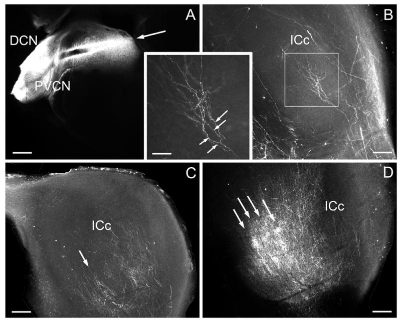
Figure 4.

Epifluorescent image showing example of DiI-pin (arrow) in the DCN in P7 ferret (A) and labeled afferent fibers ending in the contralateral IC (ICc) for a P4 (B) , P7 (C), and P14 (D) case. Boxed area in B is inset showing arborization (arrows) of cochlear nucleus fiber. Regions of dense banded labeling are indicated by arrows in C and D. A-D – calibration bars equal 200μm; inset – calibration bar equal 80 μm.
