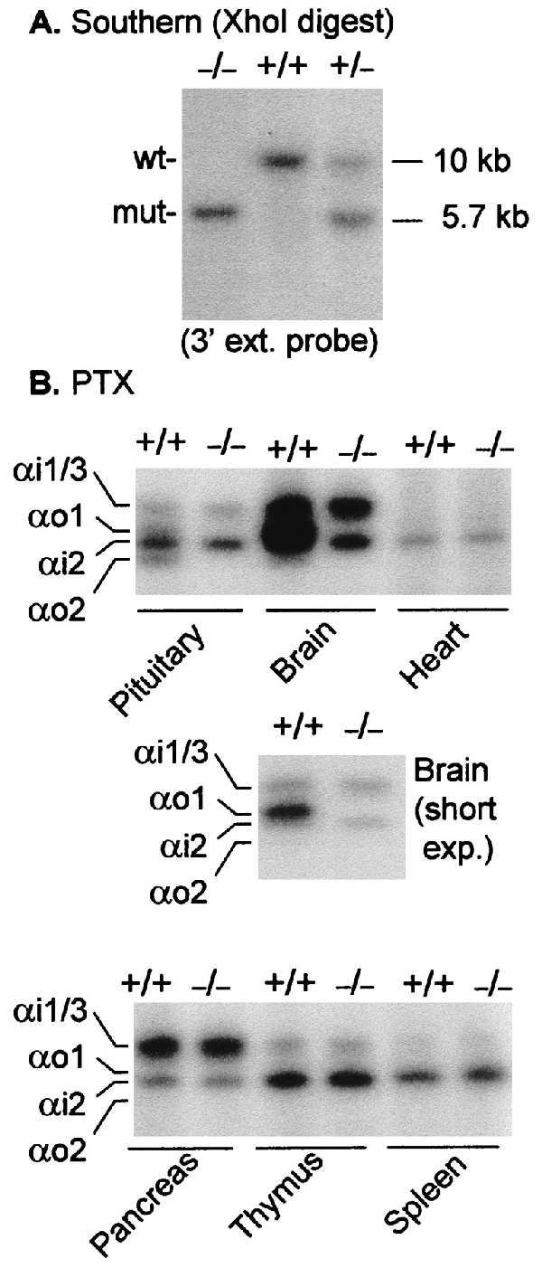
Figure 2.

Genotype and ADP-ribosylation patterns of wild-type and αo−/− mice. (A) Southern blot analysis of XhoI-digested DNA from tail biopsies of −/− mice and +/− and +/+ littermates; the probe was the 3′ external probe shown in Fig. 1. (B) Selected tissues from wild-type mice and mice predicted by Southern blot analysis to be Go-deficient. Homogenizations were all in 27% sucrose/1 mM EDTA/10 mM Tris⋅HCl, pH 7.5. Homogenates were ADP-ribosylated without further processing in the presence of guanosine 5′-[β-thio]diphosphate, adenosine 5′-[β,γ-imido]triphosphate, 0.1% SDS, and 2 mM DTT for 30 min at 32°C in a final volume of 15 μl as described (36). NAD was then added to give 4 mM, mixed with 30 μl of 2× Laemmli’s sample buffer, and separated by urea-gradient SDS/PAGE in 9% gels (36). The gels were stained and destained over night and autoradiographed. Except for brain, the predominant bands are αi2. Note that in the pituitary the intensities of αo1 and αo2 (above and below αi2) are about equivalent; in brain, αo1 is the predominant band with αi1/3 and αi2 being similar in intensity. In heart ventricle homogenates, only the αi2 band is evident [absent in αi2-deficient hearts (37)].
