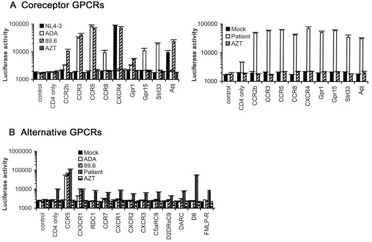
Figure 1. Coreceptor usage.
(A) Cf2-Luc cells were transfected with pcDNA3-CD4 alone or cotransfected with pcDNA3-CD4 and pcDNA3 expressing CCR2b, CCR3, CCR5, CCR8, CXCR4, Gpr1, Gpr15, Strl33 or Apj and infected with equivalent amounts of each control HIV-1 virus (left panel) or patient-derived virus (right panel). Control cells were transfected with pcDNA3 plasmid only. Mock-infected cells were treated with culture medium. Cell lysates were prepared at 48 h post-infection and assayed for luciferase activity. (B) Cf2-Luc cells transfected with pcDNA3-CD4 alone or cotransfected with pcDNA3-CD4 and plasmids expressing CCR5, CX3CR1, RDC1, CCR7, CXCR1, CXCR2, CXCR3, C5aRC9, D2DRsC9, DARC, D6 or FMLP-R were infected with equivalent amounts of each HIV-1 virus. HIV-1 entry was measured as above. Data are represented as means from duplicate infections. Error bars represent standard deviations. Similar results were obtained in two independent experiments.

