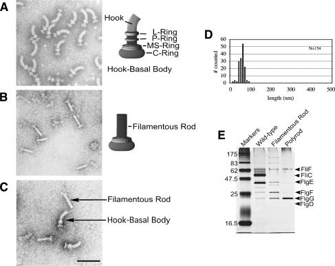
Figure 1.
The flgG regulatory mutants result in a filamentous rod phenotype. Structures isolated from flgG* regulatory mutants. For visualization by electron microscopy, cells and isolated flagellar structures were stained with 1% PTA (pH 7 or pH 5) and observed with a JEOL 1200Ex electron microscope at 80 kV. Electron micrographs of isolated HBB structures from wild-type cells (SJW1103) (A) and from the ring-defective (ΔflgHI) flgG regulatory (flgG*) double mutants (TH5931) (B), and a mixture of HBB and filamentous rod structures from strains SJW1103 and TH5931 (C). (D) The rod length measurements of isolated flagellar–basal structures from a ring mutant strain with a FlgG*-rod (TH5931). (E) SDS-PAGE analysis of protein from flagellar structures isolated from wild-type cells (SJW1103) including filaments, filamentous rod structures from strains lacking filaments (TH5931, FlgG*-rod), and polyrod structures from strains lacking filaments (TH9709, ΔfliK FlgG*-rod). The concentration of FliF in each fraction was first determined and the amount of extract loaded was based on equal amounts of FliF.

