
FIG. 4. Heat shock induces GSK-3β nuclear accumulation.
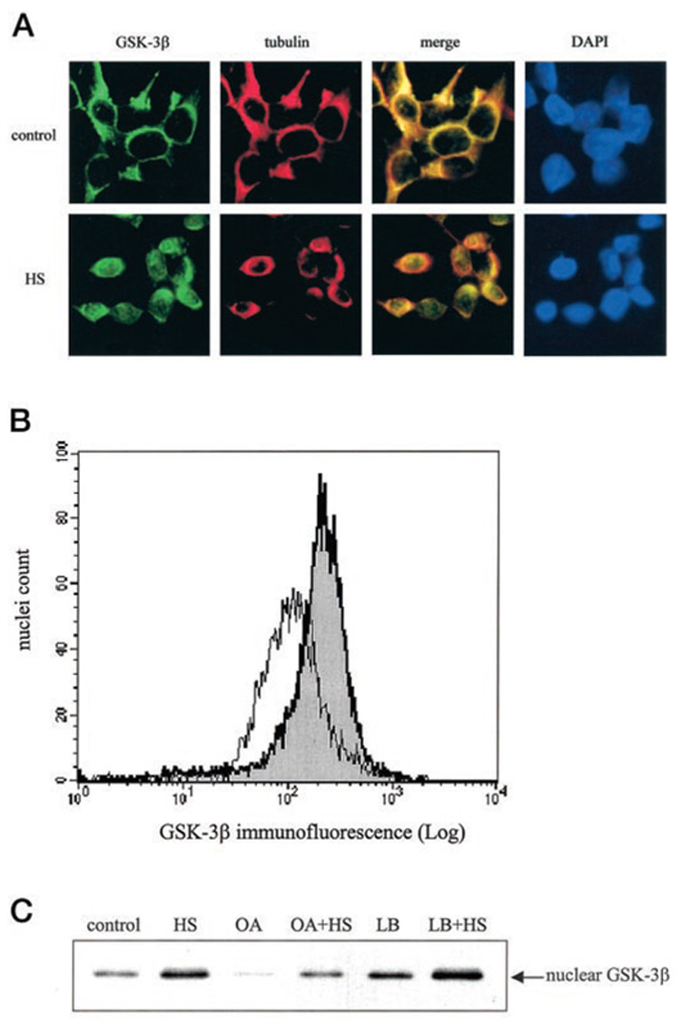
A, cells cultured on glass coverslips were either maintained in a 37 °C chamber as controls or subjected to heat shock (HS) at 45 °C for 30 min and then transferred to a 37 °C chamber for an additional 30 min. Cells were fixed, permeabilized, and immunofluorescently labeled as described under “Experimental Procedures.” Cells immunofluorescently labeled with GSK-3β-fluorescein isothiocyanate-conjugated antibody fluoresce green, and cells labeled with tubulin-Texas Red-conjugated antibody fluoresce red. Colocalization of GSK-3β and tubulin appears yellow in the merged image. Nuclei were visualized by 4,6-diamino-2-phenylindole (DAPI) staining. A representative section is shown at 400× magnification. B, isolated nuclei from control cells (white peak) and heat shock-treated cells (gray peak) were fixed, immunofluorescently stained with GSK-3β and stained with Hoechst 33342, and analyzed by flow cytometry as described under “Experimental Procedures.” C, control and heat shock-treated (HS) cells were incubated with 100 nM okadaic acid (OA) 1 h prior to heat shock treatment or with 10 ng/ml leptomycin B (LB) 2.5 h prior to heat shock treatment. The nuclear extracts were immunoblotted for GSK-3β.