
Figure 1. Dynamic cytoskeletal changes during Nasonia oogenesis.
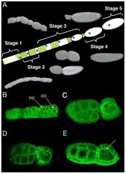
The 5 stages of oogenesis as described by King & Richards (1969). Yellow and white represents oogonium, nurse cells are in yellow, oocyte in white, oocyte nucleus in blue and vitelline membrane in grey. Photographs of fixed wild type follicles taken using Nomarsky optics. In early stage one follicles, the oocyte and nurse cells are indistinguishable. Later stage one follicles begin to distinguish the nurse cells from the oocyte which lies at the posterior of each follicle. Stage 2 follicles have the nurse cells and a smaller oocyte separated from one another with a constriction. In early stage 3 follicles, the oocyte and nurse cells are comparable in size. In later stage 3 follicles, however, the oocyte is larger than its accompanying nurse cells. Stage 4 follicles empty the nurse cell contents into the oocyte. In stage 5, a vitelline membrane is laid around the oocyte (A). Early stage 2 follicles stained using alpha tubulin antibody and analyzed using confocal microscopy. nc, nurse cells, oc, oocyte (B). Later stage 3 follicles show a complex microtubule network throughout the oocyte and nurse cells, and highly enriched at the oocyte nucleus (C,D). Late Stage 3 follicles show a breakdown of the microtubule cytoskeleton along the anterior-posterior axis, but retain a hairpin like organization surrounding the oocyte nucleus (arrowhead) (E).