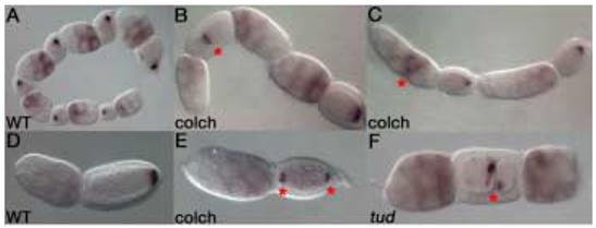
Figure 2. nos mRNA localization is microtubule independent.

Wild type nos mRNA localization (A,D). Inverted, fused follicle, marked by an asterisk, is shown on the left with posterior nos localization. The adjacent follicle on the right shows the normal orientation of the follicular chain (B). Follicles often display extra nurse cells, with nos mRNA accumulating in central nurse cells (marked by an asterisk) in response to colchicine-treatment (C). A bipolar follicle shows localization of nos to both poles of the oocyte (E). Bipolar localization is marked by asterisks. tud RNAi results in dicephalic follicles with abnormal nurse cell numbers (F). The furrow within the oocyte is marked by an asterisk. Nonetheless, tud RNAi affected follicles show normal posterior nos mRNA localization (F).
