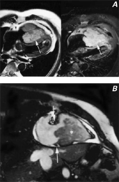
Fig. 1 A) Spin echo T2-weighted (left) and contrast-enhanced T1-weighted (right) images show a large mass (arrows) nearly filling the right ventricular cavity. The mass has lobulated borders and is adherent to and infiltrating the distal interventricular system. B) Cine magnetic resonance image obtained in the 2-chamber long-axis projection through the right ventricle. The mass (asterisk) is filling the right ventricular cavity and projecting posteriorly through the tricuspid valve (the tricuspid annulus is between the white arrows).
