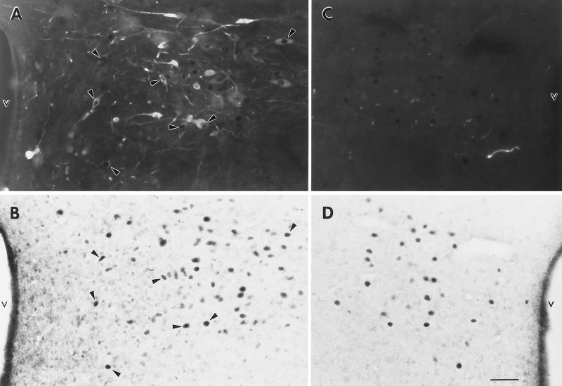
Figure 2.
Fluorescent OT-ir (A) and light-field nuclear ERβ-ir (B) in the PVN of a 40-μm-thick section at the level of approximately B −1.90. Arrowheads denote examples of dual-labeled cells (compare arrowheads in A and B). Note the absence of cell soma AVP-ir at a comparable level (C), where numerous cells containing nuclear ERβ-ir are seen (D). Photomicrographs were taken at ×200 under a Nikon light microscope and a 35 mm camera by using Kodak technical pan film. v, Third ventricle. (Bar = 40 μm.)

