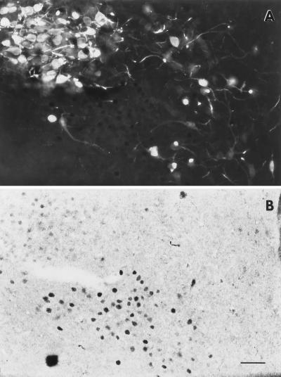
Figure 3.
Fluorescent AVP-ir (A) and light-field nuclear ERβ-ir (B) in the PVN of a 40-μm section, approximately at B −1.78. Note the very dark, well-defined nuclear ERβ-ir within the lower, central half of panel (B), which corresponds primarily to the medial parvicellular, ventral, and dorsal zones. In contrast, note the very light, yet distinct ERβ-ir in the upper left corner of B, which corresponds to the posterior magnocellular part, lateral zone. Scattered cells containing AVP-ir were found in the parvicellular regions, but only an occasional cell appeared to contain both AVP- and ERβ-ir (none demonstrated here). Most ERβ-ir within the magnocellular area appeared to be located adjacent to, but not in, the AVP-ir magnocellular neurons. Photomicrographs were taken at ×200 under a Nikon light microscope and a 35 mm camera by using Kodak technical pan film. (Bar = 40 μm.)

