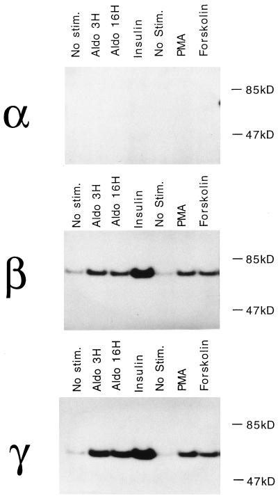
Figure 3.
Phosphorylation of ENaC after stimulation by aldosterone, insulin, and activation of kinases. αβγ MDCK cells were labeled with 32P followed by no treatment, 3 and 16 h of aldosterone (Aldo), or 15 min of insulin, PMA, or forskolin/3-isobutylmethylxanthine. α, β, and γ subunits were immunoprecipitated from equal amounts of cell lysates, and the products were resolved by SDS electrophoresis.

