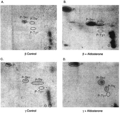
Figure 4.
Phosphoamino acid analysis of β and γ subunits of the ENaC without and with aldosterone. αβγ MDCK cells were grown in serum-free medium for 24 h before experiments. Cells were then treated with aldosterone for 16 h. During the last 3 h of incubation, 1 mCi/ml 32P was added to the medium. β and γ subunits were isolated by immunoprecipitation followed by SDS/gel electrophoresis. Acid hydrolysates of the proteins were spotted onto cellulose plates and separated by thin-layer electrophoresis in two dimensions. Middle circles indicate migration positions of phosphorylated standards; O is the origin and Pi is the completely hydrolyzed phosphate.

