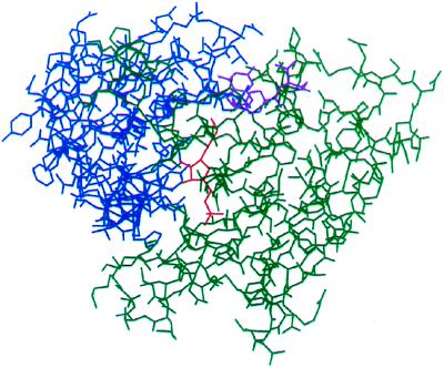
Figure 1.
Crystal structure of purN GAR transformylase complexed with β-GAR and N10-formyltetrahydrofolate. purN is shown bound to β-GAR (red) and N10-formyltetrahydrofolate (purple) and colored to show that the protein falls into two discrete domains—one responsible for binding GAR (green) and one domain that binds the N10-formyltetrahydrofolate (blue). The active site is at the interface between the two domains.

