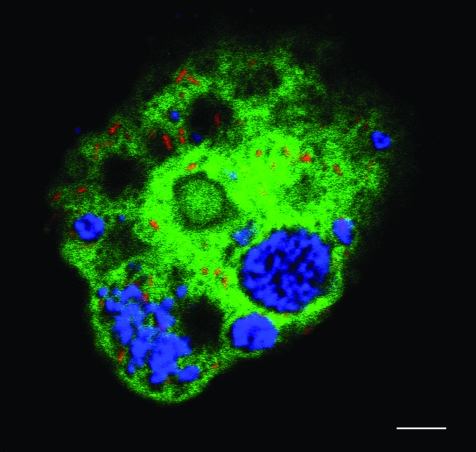
Fig. 2.
In situ identification of the two endosymbionts of Acanthamoeba sp. OEW1, Parachlamydia sp. OEW1 and ‘Cand. Procabacter sp. OEW1’. The probes used in this analysis were Proca-438 directly labelled with the hydrophilic sulfoindocyanine fluorescent dye Cy3 specific for ‘Candidatus Procabacter acanthamoebae’ (Horn et al., 2002), Bn9-658 labelled with Cy5 targeting a subgroup of the Parachlamydiaceae (Amann et al., 1997) and EUK-516 labelled with 5(6)-carboxyfluorescein-N-hydroxy-succinimide (FLUOS) targeting most members of the Eukarya (Amann et al., 1990). To ensure specificity hybridization was performed with 20% formamide in the hybridization buffer and corresponding salt concentration in the washing buffer. For further details on oligonucleotide probes, see probeBase at http://www.microbial-ecology.net/probebase (Loy et al., 2007). The overlay of the FISH micrographs, illustrating Cy3 in red, Cy5 in blue, and FLUOS in green, demonstrates the intracellular location of both endosymbionts within the same amoeba host cell. Fluorescence in situ hybridization was performed as described previously (Manz et al., 1992; Horn et al., 2001) and examined by a confocal laser scanning microscope (LSM 510 Meta, Carl Zeiss, Jena, Germany). All experiments were performed at least three times and yielded consistent results. Intervals of at least 1 week separated individual experiments. Bar, 5 μm.

