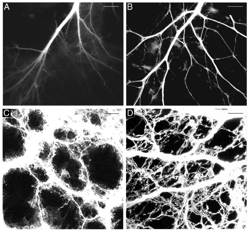
Fig. 1.

DiI-labeled vagal anterior gastric branches adjacent to the lower esophageal sphincter at P0 (A,B) and axons in the forestomach at E16.5 with axon bundles overexposed to permit visualization of individual fibers (C,D). A. A fluorescence photomicrograph illustrating the reduced visibility of labeled fibers 6 wk after application of dried DiI oil (Experiment 1). B. A confocal image illustrating the more rapid DiI diffusion, more complete labeling, and absence of DiI leakage 2.5 wk after DiI crystal application and use of EDTA in the fixative. C. Confocal image of DiI-labeled tissue mounted in 70% glycerol, 5% n-propyl gallate. D. A confocal image of DiI-labeled axons from a comparable stomach region to that shown in C, but imaged from a stomach mounted in PBS. Scale bars = 200 μm (A,B), and 25 μm (C,D).
