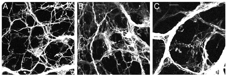
Fig. 2.

Confocal images illustrating the development of vagal axon bundles in the myenteric plexus of the forestomach. A. At E13.5 a plexus-like pattern started to emerge and axon bundles were small in diameter. B. At E14.5 axon bundle diameters and distances between axon bundles increased, the latter due to stomach growth. C. By E16.5 axon bundle diameters and interbundle distances had again increased; a putative IGLE precursor is present in the center of this image. Scale bars = 25 μm (A,B,C).
