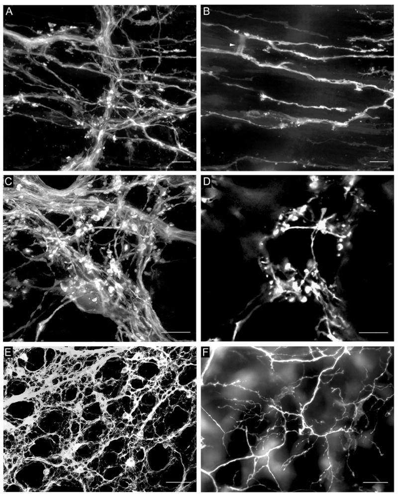
Fig. 3.

A. DiI-labeled fibers in the myenteric plexus and putative IMA precursors in the underlying circular muscle. B. The same field as in A with imaging restricted to the circular muscle layer. DiI-labeled putative IMA precursors were present with two of their telodendria connected by a cross-bridge fiber (arrowhead). C. DiI-labeled fibers in the myenteric plexus and a putative IGLE precursor immediately below the plexus. D. The same field as in C with imaging restricted to the tissue plane immediately below the myenteric plexus, which contained the putative IGLE precursor. E. DiI-labeled fiber bundles and numerous putative efferent terminals in the myenteric plexus. F. The same field as in E with only the submucosa and mucosa imaged, which contained a network of fiber bundles, single axons and nerve terminals that originated in the myenteric plexus. All images were from P0 mice. Scale bars = 10 μm (A–D) or 100 μm (E,F).
