
Fig. 6.
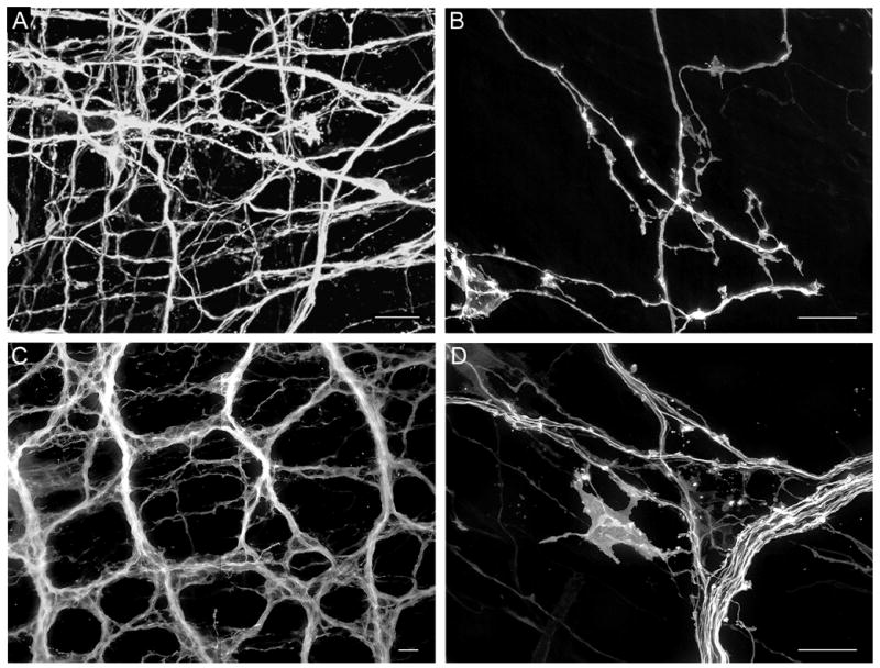
A. Vagal myenteric network of the esophagus labeled at P0. B. Individual labeled fibers with small growth-cone like structures in the esophageal myenteric plexus at P0. C. DiI-labeled myenteric fiber bundles, axons and putative efferent terminals in the duodenum at P0. D. Labeled fiber bundle, single axons and large growth-cone like structure in the duodenal myenteric plexus at P0. Scale bars = 20 μm (A – D).