
Fig. 1.
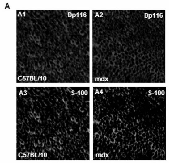
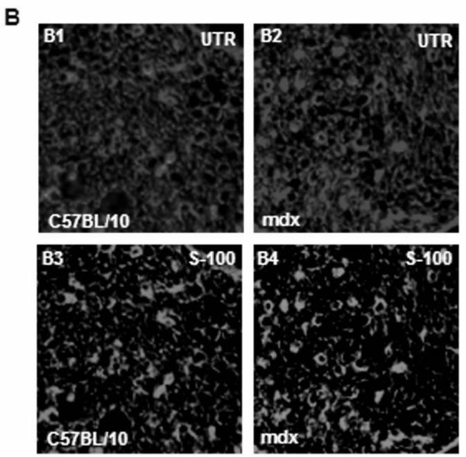
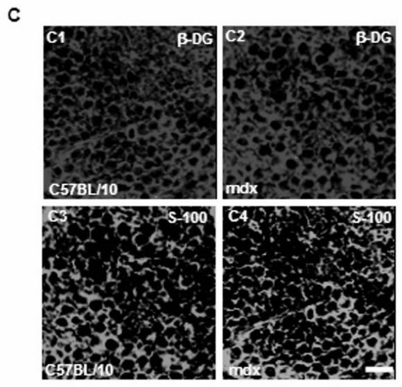
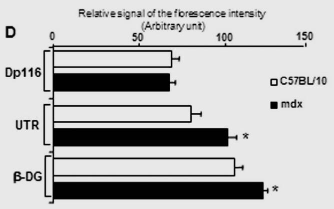
(A) Localization of DAP complex in normal (C57B/10) and mdx transversal peripheral nerve sections. Dp116 (A1, A2) and utrophin (UTR) (A3, A4) were detected in Schwann cell outer membrane as shown by the S-100 antibody (A5, A6) which labels myelinated and non-myelinated Schwann cells. (B) β-dystroglycan (β-DG) (B1, B2), α-dystrobrevin1 (α-DB1) (B3, B4), and α7B-integrin (α7B-ITG) (B5, B6) detection on transversal PN section in C57BL/10 and mdx nerve showed different localization of these proteins in nerve. β-DG which showed the most intensive labelling and α-DB1 were localized in the in Schwann cell outer membrane whereas α7B-ITG is expressed in axon region. Bar = 15 μm.