
Fig. 2.
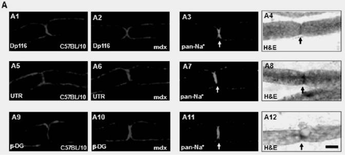
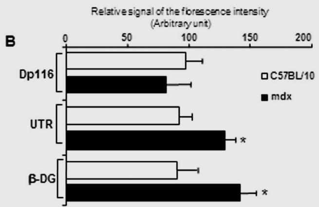
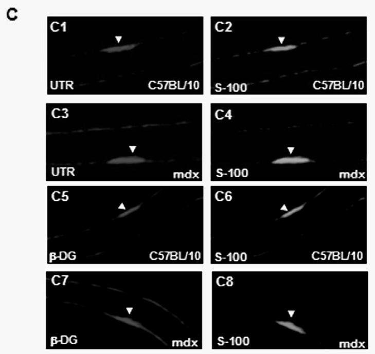
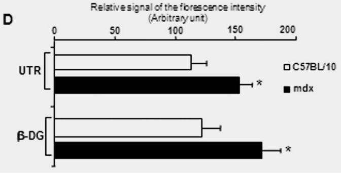
(A) Localization of Dp116 (A1, A2), Utrophin (UTR) (A5, A6) and β-dystroglycan (β-DG) (A9, A10) in nodes of Ranvier. A3, A7, A11 showed the specific labelling of Nodes of Ranvier (arrows) with the pan-Na+ antibody and the Hematoxylin/eosin (H&E) staining (A4, A8, and A12). Dp116 seems deceased in some nodes of Ranvier in mdx sections (A2), whereas utrophin and β-DG (A6 and A10 respectively) were highly detected compared with C57BL/10 (A5 and A9). (B) Immunolabelling of utrophin (UTR) (B1, B3) and β-dystroglycan (β-DG) (B5, B7) in the outer membrane of Schwann cells on isolated C57BL/10 and mdx nerve fibres. The same Schwann cells (Arrow head) were also detected by the S-100 antibody (B2, B6 and B4, B8) 5μm.