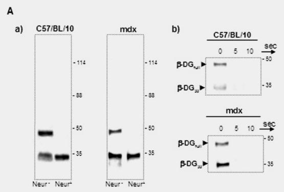
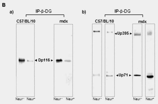
Fig. 7.


Blot overlay analysis of the binding of Dp116 and utrophin isoforms with the Schwann cell membrane protein fraction of mdx mouse. Mdx Schwann cell membrane was separated by 5–15% SDS-PAGE, transferred to nitrocellulose membrane, and overlaid O/N at 4°C with the crude membrane fraction after β-DG immunoprecipitation (IP- β-DG) (see Fig. 5). After intensive washing, the nitrocellulose membrane was revealed by the dystrophin antibody (panel a) and the utrophin antibody (panel b). In panel a, the arrows show positive bands with an Mr. of 117 kDa (corresponding to the Dp116 band) and an intensive 49 kDa band revealed with dystrophin antibody that corresponds to the Dp116 fraction associated with the 43 kDa β-DG after overlay. Panel b shows the nitrocellulose membrane revealed by utrophin antibody after overlay. Three bands were detected (70 kDa, 49 kDa and 30 kDa). The 70 kDa band corresponds to Up71, the second band corresponds to the amount of utrophin fraction which bound the β-DGfull, and the last band (most intensive) corresponds to the utrophin fraction which bound to β-DG30. Panel c represents the Western blot revelation of β-DG, utrophin, and dystrophin before overlay.
