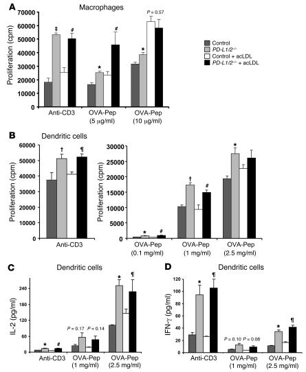
Figure 6. PD-L1/2 deficiency results in enhanced CD4+ T cell responses to APCs in vitro.
Proliferative response of OVA peptide–specific CD4+ cells were measured when cultured with wild-type C57BL/6 (Control) or PD-L1/2–/– APCs with or without lipid loading with acLDL in the presence of anti-CD3 or antigen stimuli. OVA pep, OVA peptide. (A) APCs were peritoneal macrophages (macrophage/T cell ratio, 2:1). (B) APCs were splenic DCs (DC/T cell ratio, 1:10). Incorporation of [3H]-thymidine was determined at 64 hours. (C and D) Cytokine secretion by the CD4+ cells cultured with DCs was measured in 48-hour supernatants. n = 3 per group. Data are mean ± SEM. *P < 0.05, †P < 0.01, ‡P < 0.001 versus control; ζP < 0.05, #P < 0.01 versus control plus acLDL.

