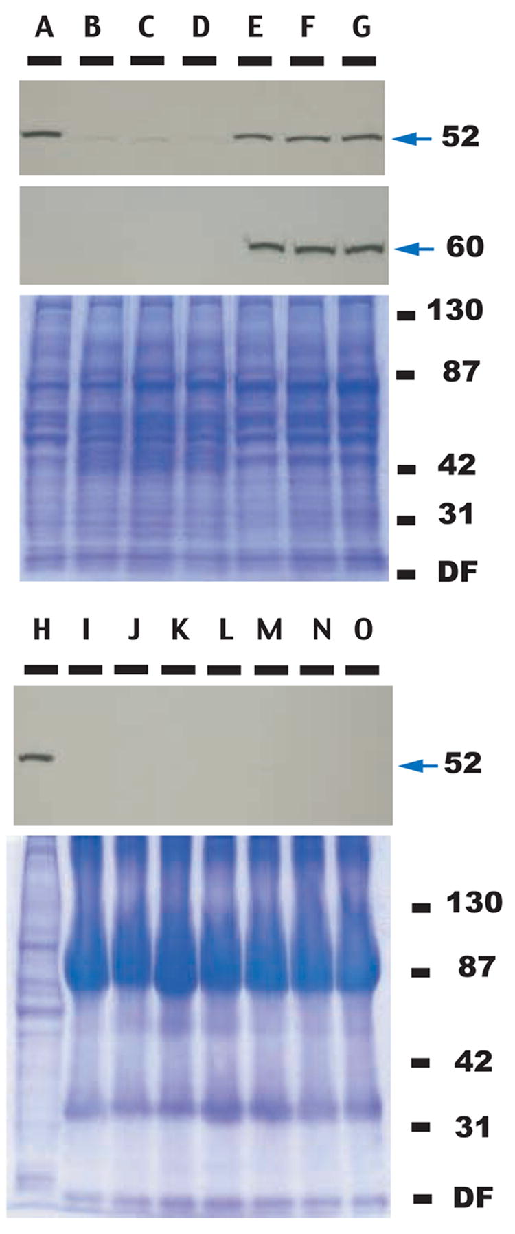
FIGURE 2. Immunoblot analysis of VIP in normal and RSV-SR transformed CEFs.

Upper three-partite panel, top panel (lanes A–G): Western blot analysis of cell lysates from CEFs (lane A), CEFs infected with RSV-SR td106 mutant (lanes B, C, D), and CEFs transformed by RSV-SR (lanes E, F, G). The blot was probed with a monoclonal antibody raised against human VIP. Lysates from cells derived from three independent experiments (infection with RSV-SR td106 mutant and transformation with RSV-SR) were used. Arrow points to VIP of 52 kDa molecular weight. Middle panel, the same Western blot was probed with src phospho-Tyr 416-specific antibody. Arrow points to c-src of 60 kDa molecular weight. Lower panel, Coomassie blue staining of the parallel SDS-polyacrylamide gel analyzed in the upper panels.
Lower two panels, top panel (lanes H–O): Western blot analysis of conditioned media from normal and RSV-SR transformed CEFs; lane H, lysate from normal, uninfected CEFs that serves as a positive control for the presence of VIP; lanes I–K, supernatant from CEFs infected with RSV-SR td 106 mutant; and lanes M–O, supernatant from CEFs transformed with RSV-SR. Arrow indicates VIP migrating at the apparent 52 kDa molecular weight. Lower panel, Coomassie blue staining of the parallel SDS-polyacrylamide gel analyzed in the upper panel.
