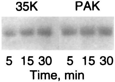
Figure 2.
Autoradiography of the heavy chain of Acanthamoeba myosin IC after phosphorylation with expressed 35K and PAK1. Myosin IC was incubated with [γ-32P]ATP and either 35K (Left) or His-PAK1 (Right) as described for the indicated times. The reactions were stopped by addition of SDS sample buffer and the samples separated by PAGE. The autoradiogram of the myosin IC heavy chains is shown. The level of phosphorylation was ≈1 mol of Pi per mol in all samples except the 5-min 35K sample, which was ≈0.8 mol of Pi per mol.

