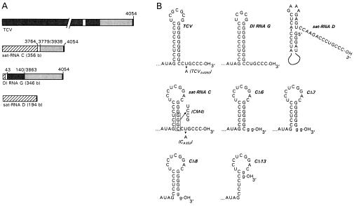
Figure 1.
Sequences and secondary structures of TCV and TCV-associated subviral RNAs. (A) Schematic representations of TCV, satellite RNA (sat-RNA) C, defective interfering (DI) RNA G, and sat-RNA D. Similar sequences are shaded alike. Positions of TCV-related sequence in the subviral RNAs are indicated. (B) Secondary structures of the 3′-terminal sequences of TCV, DI RNA G, sat-RNA D, sat-RNA C (16), and mutants derived from sat-RNA C. Plasmid-derived nucleotides present in the transcripts of sat-RNA C mutants containing 3′-terminal deletions are in lowercase letters. Point mutations that were generated in wild-type TCV and sat-RNA C are indicated, with names of mutants in parentheses.

