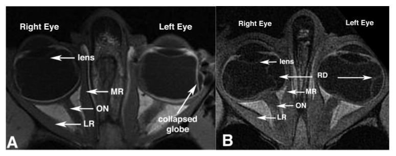
Figure 1.

Axial MRIs showed no systematic differences between the orbits of orthotropic and strabismic monkeys. Left: orthotropic M. mulatta 94D248. Resolution, 273 μm in a 3-mm-thick plane. Right: artificially induced right esotropic M. mulatta RRm5, which had undergone monocular occlusion of the right eye. Resolution, 234 μm in 2-mm-thick plane. RD, retinal detachment.
