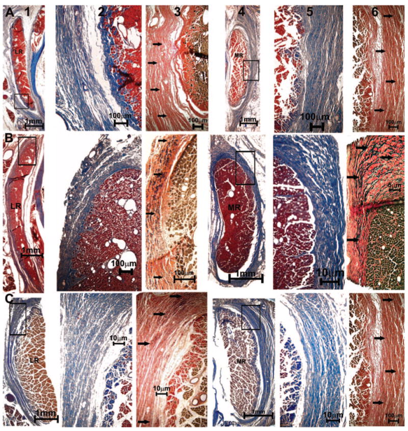
Figure 10.

Quasicoronal histologic sections of monkey orbits stained with MT and EVG stains. (A) Normal (94D248 right orbit), (B) naturally esotropic (124G left orbit), and (C) artificially esotropic (RHn4 left orbit) monkeys. Columns from left to right: lateral rectus (LR) EOM (1), magnified inset of pulley in MT stain (2), and elastin fibrils (arrows) in EVG stain (3), MR EOM (4), magnified inset of collagen fibers in MT stain (5), and elastin fibrils (arrows) in EVG stain (6).
