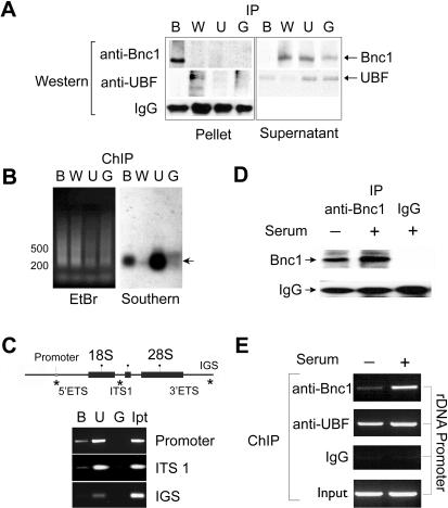
Figure 1. Basonuclin interacts with rDNA promoter in vivo.
A, Western analyses of immunoprecipitated proteins from HaCaT cell lysate. The IP antibodies are shown above the Western blot; B, anti-basonuclin (alpha-hB34), U, anti-UBF, W, anti-WT protein, and G, IgG from a naive rabbit. The detecting antibodies are indicated on the left. The target proteins were monitored in both the pellet and supernatant. Note that no cross-reactivity was observed. B, The resolution of the ChIP assay was assessed by a Southern analysis. A primer was ligated to the immunoprecipitated DNA, which was then amplified by PCR. The amplified DNA was separated by agarose electrophoresis and visualized by ethidium bromide (EtBr). The DNA was then transferred to a nylon membrane and probed with an rDNA promoter probe (Southern). The chromatin immunoprecipitation (ChIP) antibodies are indicated above the gel by letters described in (A). A DNA size marker is shown on the left. The DNA fragments detected by the probe are indicated with an arrow on the right. C, Basonuclin's association with three regions of rDNA was investigated with ChIP-PCR. The top panel depicts a generic rDNA transcription unit. Indicated are the promoters, the rRNA coding sequences (18S and 28S), the external and internal transcribed spacers (ETS and ITS) as well as the intergenic spacer (IGS). Regions tested by PCR are indicated with a (*). Twice-ChIP-precipitated DNAs were used as template for PCR, whose products were analyzed by electrophoresis (lower panel). The ChIP antibodies are indicated above the gel (B, U, G, as in A, Ip, Input DNA). PCR specificity is shown on the right. D, Basonuclin level in HaCaT cells cultured in the presence (+) and absence (-) of serum. Basonuclin was immunoprecipitated from cell lysate and analyzed by Western blot. The precipitation antibodies are indicated above the gel image and the Western detecting antibodies on the left. E, The association of basonuclin and UBF to rDNA promoter in the presence and absence of serum. ChIP-precipitated DNA was used as templates for PCR detection of the rDNA promoter. ChIP-antibodies are listed to the left of gel image.

