
Fig. 2. Methylation specific PCR (MSP), quantitative analysis: Sensitivity of detecting methylated and unmethylated HPV-18 L1 DNA with specific primers.
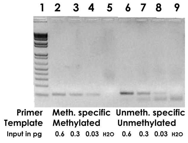
Lanes 2, 3 and 4 show the amplification of 0.6, 0.3, and 0.03 pg of Sss I methylated HPV-18 plasmid with methylation specific primers, lanes 6, 7, and 8 amplification of 0.6, 0.3, and 0.03 pg of unmethylated HPV-18 plasmid with primers specific for unmethylated DNA. The weak bands in slots 4 and 8 were the lowest sensitivity that could be achieved and correspond to about 2,500 molecules, a satisfactory detection limit in consideration of the fact that bisulfite modification fragments about 99% of the target DNA to sizes too small for PCR detection. Slot 1, size marker, slot 5 and 9, negative control (water).